

Dubai Doctors Remove 2cm-Long, 1cm-Wide Fish Bone from 60-Year-Old Man’s Airway.
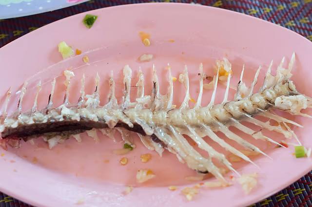
Dubai: A 60-year-old Indian expatriate living in Sharjah endured nearly two months of persistent coughing, chest congestion and worsening breathing difficulties after a sharp fish bone became lodged deep inside his airway.
The patient, Unnikrishnan Sivaraman, had accidentally inhaled the bone while eating. Measuring approximately 2cm in length and 1cm in width, the foreign object remained trapped in his respiratory passage for several weeks.
On September 11, a specialist medical team at Aster Hospital Mankhool in Dubai successfully removed the bone through a medical procedure.
According to details released by the hospital on Monday, Unnikrishnan recovered sufficiently to return home within hours of the intervention.
How a Fish Bone Led to Two Months of Breathing Problems
The incident began on July 1, when Unnikrishnan accidentally inhaled a bone from a milkfish while having a meal.
Initially, he experienced an uncomfortable sensation, but his condition later became more concerning when he developed severe nausea and vomited material containing traces of blood.
Alarmed by these symptoms, he sought emergency medical attention at Aster Hospital Al Qusais (AHQ).
However, his discomfort continued despite the initial consultation.
Over the following weeks, he experienced a persistent cough, congestion in his chest and increasing difficulty breathing.
As the symptoms failed to improve and gradually became more severe, he sought additional medical advice.
A CT scan conducted at another hospital eventually identified the underlying problem: a fish bone had become lodged in his lower respiratory tract.
Following the discovery, Unnikrishnan was referred back to Aster Hospital Al Qusais for further assessment and management.
Because the foreign object was deeply embedded and required specialised treatment, he was subsequently transferred to Aster Hospital Mankhool, where doctors could perform the necessary intervention.
Why Was the Fish Bone Difficult to Remove?
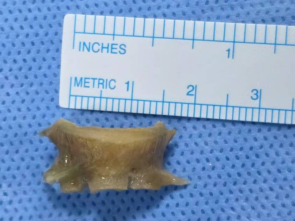
Fish bones are among the foreign objects most frequently associated with accidental ingestion or inhalation in adults, with studies reporting a higher incidence of such incidents among Asian populations.
However, Unnikrishnan’s case was particularly challenging because the bone had travelled beyond the upper airway and become lodged deep within the lower respiratory system.
The sharp fragment was trapped in the airway leading to his right lung, making its removal more complicated than a typical case involving a fish bone stuck in the throat.
Its location and the length of time it had remained inside the airway required specialist medical attention to remove it safely.
Unnikrishnan arrived at Aster Hospital Mankhool on August 26, where specialists examined his previous CT scans and conducted additional medical assessments.
The evaluation confirmed that the fish bone was still lodged in the right main bronchus, the major airway carrying air into the right lung.
Because the sharp object had remained trapped for several weeks, it had triggered considerable inflammation in the surrounding tissues, along with the development of granulation tissue around the bone.
Doctors explained that the foreign object’s size, sharp edges and deep location made the extraction particularly complicated.
The medical team had to carefully consider several potential risks, including damage to the bronchial wall and nearby structures, bleeding, sudden blockage of the airway and difficulties maintaining sufficient oxygen levels during the procedure.
These concerns made detailed preparation and close coordination between specialists essential.
Three Specialist Teams Work Together to Remove Fish Bone
A multidisciplinary team comprising specialists from Pulmonology, Ear, Nose and Throat (ENT), and Anaesthesia developed a coordinated treatment plan to safely extract the foreign object.
The doctors took into account the bone’s position, sharp edges and the inflammation caused by its prolonged presence inside the airway.
According to the hospital, Dr Jolsana Augustine, Specialist Pulmonologist, performed a bronchoscopy with anaesthesia support from Dr Abhishek Jayaprakash Narayan and Dr Siraj Madayi Meethel.
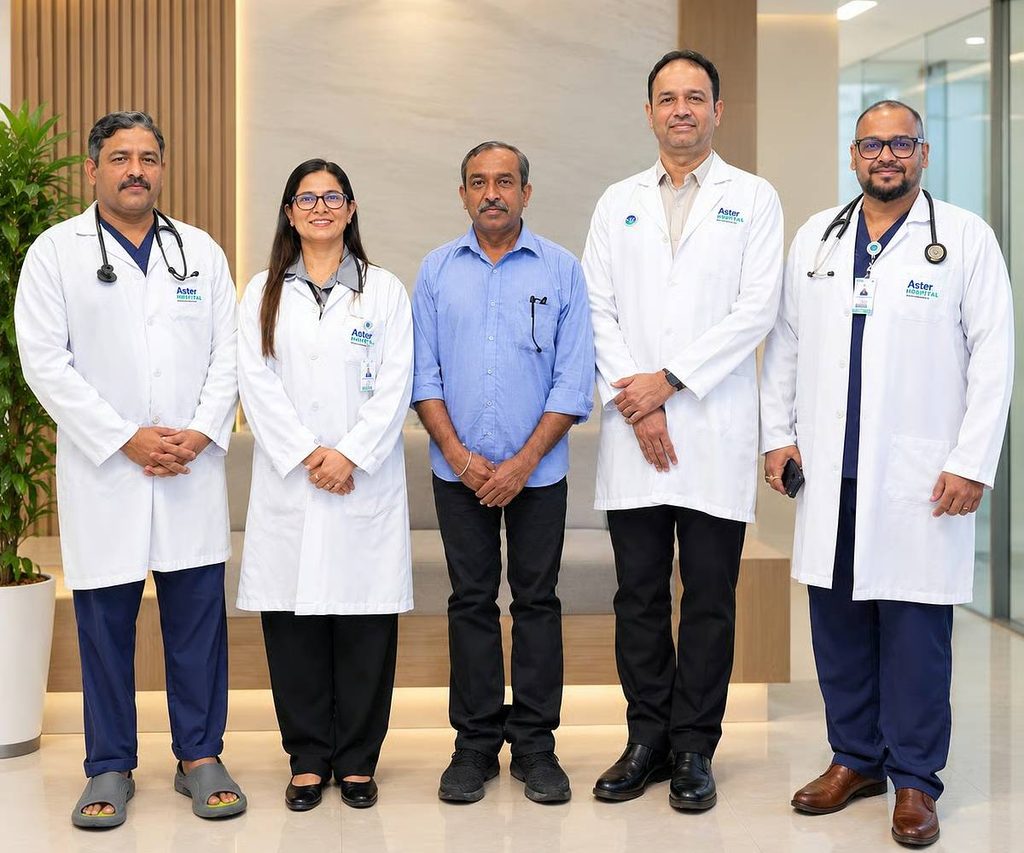
During the procedure, Dr Jolsana carefully loosened and guided the fish bone from the right main bronchus towards the upper respiratory passage.
Once the bone had been moved into a position suitable for extraction, Dr Bharath Neranki Satish Rao, Specialist ENT, performed a direct laryngoscopy.
Using specialised medical forceps, he successfully removed the sharp foreign object while taking precautions to protect the patient’s vocal cords.
Following the extraction, doctors carried out another bronchoscopy to examine the airway, assess its condition and remove accumulated secretions.
Doctors Explain the Challenges of the Procedure
Dr Jolsana Augustine said that foreign objects remaining inside the respiratory tract for extended periods can become increasingly difficult to remove because of the body’s inflammatory response.
“When a foreign body remains in the airway for a prolonged period, inflammation and granulation tissue can make removal more challenging,” she explained.
She added that the fish bone’s dimensions, sharp edges and location required a carefully planned approach to minimise the possibility of complications.
“In this case, the size, sharp edges and location of the fish bone required careful planning and controlled manipulation. Close coordination between the Pulmonology, ENT and Anaesthesia teams was essential for its safe removal while minimising the risk of airway injury or bleeding,” Dr Jolsana said.
Dr Bharath Neranki Satish Rao highlighted the delicate nature of the final extraction, particularly because the sharp bone had to pass close to the patient’s vocal cords.
“The sharp edges of the fish bone and its proximity to the vocal cords made the final stage of extraction particularly delicate,” he said.
He explained that protecting the vocal cord structures while safely removing the object was a key priority throughout the procedure.
“Our focus was to remove the foreign body safely while protecting the vocal cord structures. The successful outcome was made possible by careful coordination between the Pulmonology, ENT and Anaesthesia teams throughout the procedure,” Dr Bharath added.
Patient Returns Home Hours After Successful Procedure
The medical team successfully completed the procedure, allowing Unnikrishnan to be discharged from the hospital within just a few hours in stable condition.
During a subsequent follow-up consultation, he reported a substantial improvement in his breathing and overall health. Doctors confirmed that he was recovering well and did not require any additional medical treatment.
Relieved after nearly two months of respiratory discomfort, Unnikrishnan expressed his heartfelt appreciation to the specialists and hospital staff who helped him recover.
“I am very thankful to Dr Jolsana, Dr Bharath and the entire team at Aster Hospital Mankhool for the care and support I received,” he said.
He particularly appreciated the clear explanations and reassurance provided by Dr Jolsana throughout his treatment.
“Dr Jolsana explained everything clearly and made me feel comfortable throughout the treatment. I am very happy with my recovery and the significant improvement in my breathing and overall condition,” he added.